
Website can be closed on 12th to 14th Jan 2025 due to server maintainance work.
Experiment: Different Stages of Mitosis
Aim:
To observe and identify the different stages of mitosis in onion root tip cells using microscopy.
Materials:
1. Microscope with high-power and oil immersion objectives
2. Prepared slides of onion root tip cells
3. Coverslips
4. Kimwipes or lens paper
5. Pen and notebook for recording observations
Experimental Procedure:
1. Place a prepared slide of onion root tip cells on the microscope stage.
2. Start with low-power magnification and focus on the root tip area.
3. Switch to high-power magnification to observe individual cells in detail.
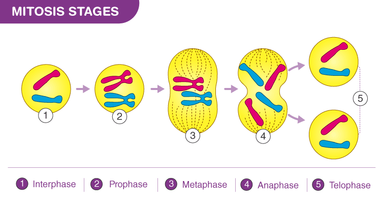
4. Identify and record the different stages of mitosis, including interphase, prophase, metaphase, anaphase, and telophase.
5. Pay attention to key characteristics of each stage, such as chromatin condensation, nuclear envelope breakdown, chromosome alignment, and sister chromatid separation.
6. Take note of the distribution and arrangement of chromosomes and microtubules during cell division.
7. Draw and label diagrams of representative cells at each mitotic stage to document your observations.
Observations:
– Interphase: Cells appear elongated with visible nuclei containing dispersed chromatin.
– Prophase: Chromatin condenses into visible chromosomes, and the nuclear envelope begins to disintegrate.
– Metaphase: Chromosomes align along the cell equator, forming the metaphase plate.
– Anaphase: Sister chromatids separate and move towards opposite poles of the cell.
– Telophase: Chromatids reach the poles, and the nuclear envelope reforms around chromatin clusters.
Results:
– Mitotic stages demonstrate distinct morphological changes indicative of cell division.
– Chromosome condensation, alignment, and segregation occur sequentially during mitosis.
– Microscopic examination allows for the visualization and identification of each mitotic stage.
Conclusion:
The observation of mitotic stages in onion root tip cells provides insights into the dynamic process of cell division. The sequential progression from interphase to telophase highlights key morphological changes associated with chromosome condensation, alignment, and segregation. Understanding mitotic stages is crucial for studying cell cycle regulation, developmental biology, and disease pathology.