
Website can be closed on 12th to 14th Jan 2025 due to server maintainance work.
Experiment: Structure of Mitochondria
Aim:
To investigate the structure and function of mitochondria through microscopic examination of cell samples.
Materials:
1. Microscope with high-power and oil immersion objectives
2. Prepared slides of various cell types (animal and plant)
3. Staining reagents (e.g., MitoTracker, DAPI)
4. Coverslips
5. Kimwipes or lens paper
6. Pen and notebook for recording observations
Experimental Procedure:
1. Prepare the microscope for use, ensuring proper illumination and focus.
2. Examine prepared slides of animal and plant cells under low-power magnification to locate mitochondria.
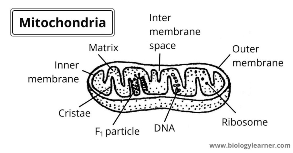
3. Switch to high-power magnification to observe the detailed structure of mitochondria, including the outer and inner membranes, cristae, matrix, and intermembrane space.
4. Record observations of mitochondrial structure, noting differences between mitochondria in different cell types and developmental stages.
5. Apply staining reagents (e.g., MitoTracker for live mitochondria, DAPI for DNA) to visualize mitochondrial components and DNA.
6. Examine the stained slides under high-power magnification to observe the distribution and morphology of mitochondria and mitochondrial DNA.
7. Discuss the functional significance of mitochondrial structures in cellular processes such as aerobic respiration, ATP production, and calcium homeostasis.
8. Consider the relationship between mitochondrial structure and cellular health, as well as the impact of mitochondrial dysfunction on human diseases.
Observations:
– Mitochondria appear as elongated organelles within animal and plant cells under high-power magnification.
– They exhibit a double membrane structure, comprising an outer membrane and inner membrane.
– Internal structures include cristae, which are invaginations of the inner membrane, and a matrix containing enzymes for ATP production.
– Staining with MitoTracker reveals live mitochondria, while DAPI staining highlights mitochondrial DNA.
Results:
– Mitochondria display a characteristic structure essential for aerobic respiration and ATP synthesis.
– Cristae provide a large surface area for ATP production through oxidative phosphorylation.
– Mitochondrial DNA is localized within the matrix, contributing to genetic control of mitochondrial functions.
Conclusion:
The microscopic examination elucidates the structure and function of mitochondria in cellular metabolism. The observed mitochondrial morphology, including membranes, cristae, and DNA, underscores their crucial role in energy production and cellular homeostasis. Understanding mitochondrial structure enhances our comprehension of cellular processes and their implications for human health and disease.