
Website can be closed on 12th to 14th Jan 2025 due to server maintainance work.
Experiment: Microscopic Examination of Nuclear Structure**
Aim:
To explore the structure and function of the nucleus through microscopic examination of prepared slides.
Materials:
1. Microscope with high-power and oil immersion objectives
2. Prepared slides of various cell types (animal and plant)
3. Staining reagents (e.g., hematoxylin and eosin)
4. Coverslips
5. Kimwipes or lens paper
6. Pen and notebook for recording observations
Experimental Procedure:
1. Prepare the microscope for use, ensuring proper illumination and focus.
2. Examine prepared slides of animal and plant cells under low-power magnification to locate nuclei.
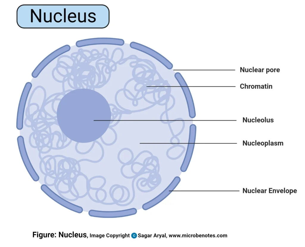
3. Switch to high-power magnification to observe the detailed structure of the nucleus, including the nuclear envelope, nucleoplasm, nucleolus, and chromatin.
4. Record observations of nuclear structure, noting differences between animal and plant cells if present.
5. Apply staining reagents (e.g., hematoxylin for nuclei) to enhance contrast and visibility of nuclear components.
6. Examine the stained slides under high-power magnification to observe the distribution and morphology of chromatin within the nucleus.
7. Note any variations in nuclear structure among different cell types and developmental stages.
8. Discuss the functional significance of nuclear components in cellular processes such as DNA replication, transcription, and regulation of gene expression.
Observations:
– The nucleus appears as a prominent structure within animal and plant cells under high-power magnification.
– It is surrounded by a nuclear envelope, enclosing the nucleoplasm and nucleolus.
– Chromatin fibers are visible within the nucleoplasm, with varying degrees of condensation.
– Staining with hematoxylin enhances the visibility of nuclear components, facilitating detailed observation of chromatin distribution.
Results:
– The nucleus exhibits a characteristic structure, including the nuclear envelope, nucleoplasm, nucleolus, and chromatin.
– Chromatin fibers appear as dispersed threads or condensed clumps within the nucleoplasm, reflecting different stages of DNA packaging and transcriptional activity.
Conclusion:
The microscopic examination highlights the nucleus’s intricate structure and pivotal role in cellular function. Understanding nuclear components and their organization provides insights into fundamental cellular processes. Further research on nuclear structure contributes to advancements in cellular biology and human health.